相關商品
商品簡介
作者簡介
名人/編輯推薦
目次
商品簡介
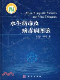
《水生病毒及病毒病圖鑒》水生病毒學研究的對象是感染水生生物或存在于水環境中的病毒。《水生病毒及病毒病圖鑒》源自著者相關研究的積累,兼具專著與圖籍屬性。從逾萬幅圖中遴選了約800幅(圖或圖版),其中彩圖約200幅。《水生病毒及病毒病圖鑒》分為6個部分:水生病毒病症及水生病毒學技術、水生哺乳動物病毒、爬行動物病毒、兩棲動物病毒、魚蝦類動物病毒及淡水浮游病毒,共分23章,以圖示和簡潔的中英文圖注直觀展示了水生生物病毒病症狀、宿主組織及其細胞的病理變化、水生病毒的超微形態及基因組成和蛋白定位等。.
作者簡介
張奇亞,中國科學院水生生物研究所二級研究員,博士生導師,魚類生物學及漁業生物技術研究中心主任、水生生物研究所學位委員會副主任;為國際病毒分類委員會(ICTV)專家組成員。主要從事水生病毒及其分子生物學研究。先後主持和承擔了國家“973”計劃、“863”計劃、國家自然科學基金和院、部、省科學基金等多個科研項目。發表研究論文150篇,其中在JournalofVirology、JournalofGeneralVirology、Virology、VirusResearch、Apoptosis、BMCgenetics、PlosOne等國際期刊上發表SCI刊源論文60篇。主編研究生教材1部,副主編或合著5部。獲授權國家發明專利5項,湖北省自然科學獎二等獎1項,中國科學院教學成果二等獎1項;獲國務院政府特殊津貼;獲中國科學院優秀教師”、湖北省“巾幗建功標兵”和“職業女標兵”等稱號。桂建芳,中國科學院水生生物研究所二級研究員,博士生導師。1994年獲首屆國家傑出青年科學基金資助;1999年9月至2007年11月任中國科學院水生生物研究所常務副所長、所長;2001年11月至2011年11月任淡水生態與生物技術國家重點實驗室主任;2004年起為國家“973”計劃項目首席科學家。已在包括Nature、PNAS、PNAS、MBE、DB、JI、MCE、BR、JournalofVirology等國際刊物發表SCI刊源論文150多篇;出版論著6部;獲獎成果7項,包括國家自然科學獎二等獎1項(2011年),湖北省自然科學獎一等獎1項(2003年),湖北省科技進步獎一等獎1項2011年);授權專利10項;培育水產養殖新品種2個。曾先後獲中國科協“青年科技獎”、中國科學院“青年科學家獎”一等獎、香港求是科技基金會傑出青年學者獎”;獲中國科學院“有突出貢獻的中青年專家”、中國科學院優秀研究生導師、湖北省優秀科技工作者、湖北省勞動模範、中華人民共和國科學技術部(簡稱科學技術部)國家重點實驗室計劃先進個人、全國“五一勞動獎章”,科學技術部“十一五”國家科技計劃執行突出貢獻獎等榮譽稱號。.
名人/編輯推薦
《水生病毒及病毒病圖鑒》是為相關學科的學生和研究者所著,可供從事病毒學、水生生物學和環境科學等相關學科教學與研究的大專院校及科研單位圖書館收藏。
目次
前言第一部分 水生病毒病症及水生病毒學技術第一章 水生生物病毒病症狀1.1 江豚皰疹樣病毒病1.2 中華鱉病毒病1.3 沼澤綠牛蛙病毒病1.4 大鯢疑似病毒病1.5 中華鱘病毒病1.6 胭脂魚病毒病1.7 長吻鮠病毒病1.8 斑點叉尾鮰病毒病1.9 鯉病毒病1.10 牙鮃病毒病1.11 草魚病毒病1.12 鱖病毒病1.13 加州鱸病毒病1.14 黃鱔病毒病1.15 大菱鮃病毒病1.16 黃顙魚疑似病毒病1.17 凡納濱對蝦病毒病1.18 近江牡蠣疑似病毒病1.19 浮絲藻病毒病1.20 魚腥藻病毒病1.21 銅綠微囊藻病毒病第二章 水生動物的細胞與組織培養2.1 內臟組織原代培養2.2 魚類皮膚組織原代培養2.3 細胞傳代培養2.4 新建細胞系核型分析2.5 細胞鹼性磷酸酶染色2.6 掃描電子顯微鏡觀察第三章 魚類病毒單克隆抗體的製備與應用3.1 細胞融合3.2 雜交瘤細胞的篩選3.3 免疫印跡分析3.4 單克隆抗體與流式細胞術3.5 單克隆抗體免疫組化檢測3.6 單克隆抗體免疫熒光觀察第四章 水生病毒的檢測4.1 細胞化學檢測4.2 組織化學檢測4.3 細胞集落形成測驗4.4 病毒侵染後細胞骨架結構的變化4.5 RNA干擾調節病毒基因的表達及其熒光檢測4.6 病毒基因產物與細胞器在宿主細胞中的共定位4.7 免疫擴散檢驗與酶聯免疫檢測4.8 流式細胞計量檢測4.9 電泳圖譜分析4.10 水生病毒蛋白質高級結構的預測4.11 水生病毒基因組組織結構4.12 水生病毒的基因功能解析4.13 含EGFP重組病毒的構建及其細胞病變空斑的識別4.14 噬藻體的檢測第二部分 水生哺乳動物病毒第五章 江豚皰疹樣病毒第三部分 爬行動物病毒第六章 中華鱉病毒6.1 超薄切片的透射電子顯微鏡觀察6.2 負染電鏡圖第四部分 兩棲動物病毒第七章 蛙虹彩病毒7.1 細胞病變7.2 病毒加工廠(病毒基質)形成7.3 病毒裝配7.4 空衣殼及異常病毒顆粒7.5 病毒在宿主細胞中的分佈7.6 病毒釋放7.7 免疫電子顯微鏡觀察7.8 純化的蛙虹彩病毒負染電鏡圖第八章 大鯢病毒樣顆粒第五部分 魚蝦類動物病毒第九章 中華鱘病毒樣顆粒第十章 胭脂魚彈狀病毒10.1 細胞感染10.2 包涵體第十一章 牙鮃淋巴囊腫病毒與正粘樣病毒顆粒11.1 淋巴囊腫病毒中國分離株11.2 牙鮃正粘樣病毒第十二章 牙鮃彈狀病毒和雙RNA病毒12.1 牙鮃彈狀病毒12.2 牙鮃雙RNA病毒第十三章 草魚呼腸孤病毒13.1 超薄切片的透射電子顯微鏡觀察13.2 負染電鏡圖第十四章 鱖球形病毒與鱖彈狀病毒14.1 鱖球形病毒14.2 鱖彈狀病毒14.3 負染電鏡圖第十五章 加州鱸呼腸孤病毒15.1 超薄切片的透射電子顯微鏡觀察15.2 負染電鏡圖第十六章 鯉春病毒血症病毒第十七章 黃鱔球形病毒和彈狀病毒17.1 黃鱔球形病毒17.2 黃鱔彈狀病毒第十八章 大菱鮃彈狀病毒和呼腸孤病毒18.1 大菱鮃彈狀病毒18.2 大菱鮃呼腸孤病毒第十九章 對蝦杆狀病毒第六部分 淡水浮游病毒第二十章 浮游病毒的多樣性20.1 淡水環境中的病毒和其他微生物20.2 長尾病毒科的噬藻(菌)體20.3 肌尾病毒科的噬藻(菌)體20.4 短尾病毒科的噬藻(菌)體20.5 球形病毒20.6 脂毛或絲杆狀病毒樣顆粒第二十一章 東湖分離的浮絲藻病毒21.1 超薄切片透射電子顯微鏡觀察21.2 負染電鏡圖第二十二章 魚腥藻病毒樣顆粒22.1 魚腥藻球形病毒樣顆粒22.2 魚腥藻雙生病毒樣顆粒第二十三章 銅綠微囊藻球形病毒樣顆粒和有尾噬藻體23.1 銅綠微囊藻球形病毒樣顆粒23.2 銅綠微囊藻有尾噬藻體超薄切片電鏡圖23.3 銅綠微囊藻有尾噬藻體負染電鏡圖參考文獻ContentsPrefaceSection 1 Viral disease symptoms of aquatic organisms and techniques in aquatic virologyChapter 1 Viral disease symptoms in several aquatic organisms1.1 Finless porpoises herpes-like virus disease1.2 Soft-shell turtle viral disease1.3 Pig frog viral disease1.4 Chinese giant salamander virus-like disease1.5 Chinese sturgeon viral disease1.6 Chinese sucker fish viral disease1.7 Chinese longnose catfish viral disease1.8 Channel catfish viral disease1.9 Common carp viral disease1.10 Flounder viral disease1.11 Grass carp viral disease1.12 Mandarin fish viral disease1.13 Perch viral disease1.14 Rice field eel viral disease1.15 Turbot viral disease1.16 Yellow catfish virus-like disease1.17 White leg shrimp viral disease1.18 Jinjiang oyster virus-like disease1.19 Filamentous cyanobacterium viral disease1.20 Anabaena viral disease 1.21 Microcystis viral disease Chapter 2 Cell and tissue culture in aquatic animals2.1 Primary culture of organs and tissues 2.2 Primary culture of fish skin tissues2.3 Cell subculture2.4 Karyotype analysis of newly established cell lines2.5 Alkaline phosphatase staining of cells2.6 Scanning electron microscopeChapter 3 Preparation and application of monoclonal antibodies against fish viruses3.1 Cell fusion3.2 Hybridoma screening3.3 Western blot analysis3.4 Monoclonal antibodies and flow cytometry3.5 Monoclonal antibodies in immunohistochemistry3.6 Immunofluorescence with monoclonal antibodiesChapter 4 Detection of aquatic viruses4.1 Cytochemistry detection4.2 Histochemistry detection 4.3 Foci-forming assay4.4 Cytoskeleton structure changes after virus infection4.5 RNA interference regulates viral gene expression and fluorescent detection 4.6 Colocalization of viral gene products and cellular organelles in host cells 4.7 Immunodiffusion test and enzyme linked immunosorbent assay 4.8 Flow cytometry detection4.9 Electrophoresis pattern analysis4.10 Prediction of advanced structures about aquatic virus proteins 4.11 Genome organization structures of aquatic viruses 4.12 Gene function analysis of aquatic viruses 4.13 Construct of recombinant virus with EGFP and recognition of cytopathogenic plaques4.14 Detection of cyanophagesSection 2 Viruses in aquatic mammalsChapter 5 Finless porpoise herpes-like virusSection 3 Viruses in reptilesChapter 6 Trionyx sinensis virus6.1 Transmission electron microscope observation of ultra-thinsections6.2 Negatively stained electron micrographSection 4 Viruses in amphibiansChapter 7 Rana grylio virus7.1 Cytopathological changes7.2 Formation of virus factory (viromatrix)7.3 Virus assembly7.4 Empty capsids and abnormal virus particles7.5 Distribution of virus particles in host cells7.6 Virus release7.7 Immune electron microscopy observations7.8 Negatively stained electron micrographs of the purified Rana grylio virusChapter 8 Chinese giant salamander virus-like particleSection 5 Viruses in fish and shrimpChapter 9 Chinese sturgeon virus-like particlesChapter 10 Chinese sucker rhabdovirus10.1 Cell infection10.2 Inclusion bodiesChapter 11 Flounder lymphocystis disease virus and orthomyxo-like virus particle11.1 Lymphocystis virus isolated from China11.2 Orthomyxo-like virus of flounderChapter 12 Paralichthys olivaceus rhabdovirus and birnavirus12.1 Paralichthys olivaceus rhabdovirus12.2 Paralichthys olivaceus birnavirusChapter 13 Grass carp reovirus13.1 Transmission electron microscope observations of ultrathin sections13.2 Negative staining electron micrographsChapter 14 Mandarin fish spherical virus and rhabdovirus14.1 Siniperca chuatsi spherical virus14.2 Siniperca chuatsi rhabdovirus14.3 Negatively stained electron micrographsChapter 15 Micropterus salmonides reovirus15.1 Transmission electron microscope observation of ultrathin sections15.2 Negatively stained electron micrographsChapter 16 Spring viremia of carp virusChapter 17 Monopterus albus spherical virus and rhabdovirus17.1 Monopterus albus spherical virus17.2 Monopterus albus rhabdovirusChapter 18 Turbot rhabdovirus and reovirus18.1 Scophthalmus maximus rhabdovirus18.2 Scophthalmus maximus reovirusChapter 19 Shrimp baculovirusSection 6 Freshwater virioplanktonChapter 20 Diversity of virioplankton20.1 Viruses and other microorganisms in freshwater environment20.2 Cyanophages (phages) in Siphoviridae20.3 Cyanophages (phages) in Myoviridae20.4 Cyanophages (phages) in Podoviridae20.5 Spherical viruses20.6 Lipothrix-or filamentous virus-like particlesChapter 21 Planktothrix agardhii virus isolated from Lake Donghu 21.1 Transmission electron microscope observation of ultrathin sections21.2 Negatively stained electron micrographsChapter 22 Anabaena spirioides virus-like particles 22.1 Anabaena spirioides spherical virus-like particle 22.2 Anabaena spirioides geminivirus-like particleChapter 23 Microcystis aeruginosa spherical virus-like particle and tailed-cyanophage23.1 Microcystis aeruginosa spherical virus-like particle23.2 Electron micrographs of ultrathin sections of Microcystis aeruginosa tailed cyanophage23.3 Negatively stained electron micrographs of Microcystis aeruginosa tailed cyanophageReferences.
主題書展
更多
主題書展
更多書展本週66折
您曾經瀏覽過的商品
購物須知
大陸出版品因裝訂品質及貨運條件與台灣出版品落差甚大,除封面破損、內頁脫落等較嚴重的狀態,其餘商品將正常出貨。
特別提醒:部分書籍附贈之內容(如音頻mp3或影片dvd等)已無實體光碟提供,需以QR CODE 連結至當地網站註冊“並通過驗證程序”,方可下載使用。
無現貨庫存之簡體書,將向海外調貨:
海外有庫存之書籍,等候約45個工作天;
海外無庫存之書籍,平均作業時間約60個工作天,然不保證確定可調到貨,尚請見諒。
為了保護您的權益,「三民網路書店」提供會員七日商品鑑賞期(收到商品為起始日)。
若要辦理退貨,請在商品鑑賞期內寄回,且商品必須是全新狀態與完整包裝(商品、附件、發票、隨貨贈品等)否則恕不接受退貨。


