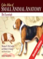
Color Atlas Of Small Animal Anatomy: The Essentials
商品資訊
ISBN13:9780813816081
出版社:John Wiley & Sons Inc
作者:Mccracken
出版日:2009/01/30
裝訂/頁數:平裝/160頁
規格:22.9cm*15.2cm*1.3cm (高/寬/厚)
版次:1
商品簡介
作者簡介
目次
相關商品
商品簡介
This new resource provides a basic foundation in small animal anatomy for students of veterinary medicine, animal science, and veterinary technology. Extraordinary accuracy and beautiful original artwork make this a truly unique learning tool that includes the anatomy of all organ systems in the dog, cat, rabbit, rat, and guinea pig - all described in a consistent manner.
Learning features include: carefully selected labeling helps students learn and remember structures and relationships; male and female of species are depicted on facing pages so topographic anatomy can be compared; structures common to various animals are labeled several times, whereas unique structures are labeled on one or two species so students can make rapid distinctions of the structures peculiar to certain animals; and an introduction that provides readers with a background in nomenclature and anatomic orientation so they can benefit from the atlas even if they lack training in anatomy.
The Atlas depicts topographic relationships of major organs in a simple, yet technically accurate presentation that's free from extraneous material so that those using the atlas can concentrate on the essential aspects of anatomy. It will be an invaluable resource for veterinary students, teachers and practitioners alike.
作者簡介
Thomas O. McCracken, MS, is Professor of Anatomy & Physiology at Robert Ross International University of Nursing (IUON) in Basseterre, St Kitts, West Indies; and Former Associate Professor of Anatomy at the College of Veterinary Medicine and Biomedical Sciences, Colorado State University.
Robert A. Kainer, DVM, MS, is Professor Emeritus of Anatomy at the College of Veterinary Medicine and Biomedical Sciences, Colorado State University, Fort Collins, Colorado.
Robert A. Kainer, DVM, MS, is Professor Emeritus of Anatomy at the College of Veterinary Medicine and Biomedical Sciences, Colorado State University, Fort Collins, Colorado.
目次
Section 1.
The Dog.
Plate 1.1 Lateral view of the dog (Beagle).
Plate 1.2 Lateral view of the bitch (Retriever).
Plate 1.3 Body regions.
Plate 1.4 Skeleton.
Plate 1.5 Cutaneous muscles and major fasciae the dog.
Plate 1.6 Superficial muscles of the bitch.
Plate 1.7 Deep muscles of the dog.
Plate 1.8 Deep cervical muscles, major joints, and in situ viscera of the bitch.
Plate 1.9 Paraxial view of the third digit.
Plate 1.10 Palmar views of the major structures of the forepaw; plantar view of the major structures of the hidpaw.
Plate 1.11 Median section of the head, and dentition.
Plate 1.12 The eye and accessory ocular structures.
Plate 1.13 The nose.
Plate 1.14 The ear.
Plate 1.15 Mouth and tongue and esophagus.
Plate 1.16 Ventral view of the abdomen and its structures.
Plate 1.17 Large intestine, anus and anal sacs.
Plate 1.18 Body cavities and serous membranes.
Plate 1.19 Thoracic, abdominal and pelvic viscera related to the skeleton of the dog.
Plate 1.20 Thoracic, abdominal and pelvic viscera, and mammary glands of the bitch.
Plate 1.21 Hip joint.
Plate 1.22 Location of major endocrine organs.
Plate 1.23 Relations of the reproductive organs of the dog.
Plate 1.24 Relations of the reproductive organs of the bitch.
Plate 1.25 Major veins.
Plate 1.26 Major arteries.
Plate 1.27 Lymph nodes and vessels.
Plate 1.28 Central and somatic nervous system.
Plate 1.29 Autonomic nervous system.
Plate 1.30 Brain, dorsal, ventral and lateral views.
Section 2.
The Cat.
Plate 2.1 Lateral view of the male cat (Moggie-nonpedigree).
Plate 2.2 Lateral view of the female cat (Persian).
Plate 2.3 Endocrine organs and lymph nodes.
Plate 2.4 Skeleton.
Plate 2.5 Cutaneous muscles and major fasciae of the male.
Plate 2.6 Superficial muscles of the female.
Plate 2.7 Middle muscles and in situ viscera of the male.
Plate 2.8 Deep muscles and in situ viscera of the female.
Plate 2.9 Median section of the head, and dentition.
Plate 2.10 Oral cavity, tongue, pharynx and esophagus.
Plate 2.11 The external, middle, and inter ear.
Plate 2.12 The eye and accessory ocular structures.
Plate 2.13 Isolated stomach and intestines.
Plate 2.14 Large intestine, anus and anal sacs.
Plate 2.15 Superficial and deep structures of the paw (foot) lateral view.
Plate 2.16 Plantar views of the major structures of forepaw and hindpaw.
Plate 2.17 Thoracic, abdominal and pelvic viscera related to the skeleton of the male.
Plate 2.18 Thoracic, abdominal and pelvic viscera, related to the skeleton of the female.
Plate 2.19 Relations of the reproductive organs of the male.
Plate 2.20 Relations of the reproductive organs of the female.
Plate 2.21 Major veins.
Plate 2.22 Major arteries.
Plate 2.23 Central and peripheral nervous system.
Plate 2.24 Brain, dorsal, ventral and lateral views.
Section 3.
The Rabbit.
Plate 3.1 Lateral view.
Plate 3.2 Body regions.
Plate 3.3 Skeleton.
Plate 3.4 Endocrine organs and lymph nodes.
Plate 3.5 Superficial muscles of the male.
Plate 3.6 Deep muscles of the female.
Plate 3.7 Median section of the rabbit’s head and dentition.
Plate 3.8 Oral cavity, tongue, pharynx and esophagus.
Plate 3.9 Thoracic, abdominal and pelvic viscera (in situ) of the male.
Plate 3.10 Thoracic, abdominal and pelvic viscera (in situ) of the female.
Plate 3.11 Relations of the reproductive organs of the male.
Plate 3.12 Relations of the reproductive organs of the female.
Plate 3.13 Central and peripheral nervous system.
Plate 3.14 Brain, dorsal, ventral, and lateral views.
Section 4.
The Rat.
Plate 4.1 Lateral view.
Plate 4.2 Skeleton of the rat.
Plate 4.3 Superficial muscles of the male.
Plate 4.4 Deep and middle muscles of the female.
Plate 4.5 Median section of the head and dentition.
Plate 4.6 Ventral view of abdominal structures (in situ) and diagram of digestive system.
Plate 4.7 Thoracic, abdominal and pelvic viscera related to the skeleton of the male.
Plate 4.8 Thoracic, abdominal and pelvic viscera, related to the skeleton of the female.
Plate 4.9 Relations of the reproductive organs of the male.
Plate 4.10 Relations of the reproductive organs of the female.
Plate 4.11 Spinal nerves.
Plate 4.12 Autonomic nerves.
Plate 4.13 Brain, dorsal, ventral, and lateral views.
Plate 4.14 Brian, sagittal section, and detail of midbrain.
Section 5.
The Guinea Pig.
Plate 5.1 Lateral view.
Plate 5.2 Skeleton.
Plate 5.3 Superficial muscles of the male.
Plate 5.4 Deep and middle muscles of the female.
Plate 5.5 Median section of the head and dentition.
Plate 5.6 Ventral view of abdominal structures (in situ) and diagram of digestive system.
Plate 5.7 Thoracic, abdominal and pelvic viscera related to the skeleton of the male.
Plate 5.8 Thoracic, abdominal and pelvic viscera, and mammary glands of the female.
Plate 5.9 Relations of the reproductive organs of the male.
Plate 5.10 Relations of the reproductive organs of the female.
Plate 5.11 Central and peripheral nervous system.
Plate 5.12 Brain dorsal, ventral, and lateral views
主題書展
更多
主題書展
更多書展今日66折
您曾經瀏覽過的商品
購物須知
外文書商品之書封,為出版社提供之樣本。實際出貨商品,以出版社所提供之現有版本為主。部份書籍,因出版社供應狀況特殊,匯率將依實際狀況做調整。
無庫存之商品,在您完成訂單程序之後,將以空運的方式為你下單調貨。為了縮短等待的時間,建議您將外文書與其他商品分開下單,以獲得最快的取貨速度,平均調貨時間為1~2個月。
為了保護您的權益,「三民網路書店」提供會員七日商品鑑賞期(收到商品為起始日)。
若要辦理退貨,請在商品鑑賞期內寄回,且商品必須是全新狀態與完整包裝(商品、附件、發票、隨貨贈品等)否則恕不接受退貨。


