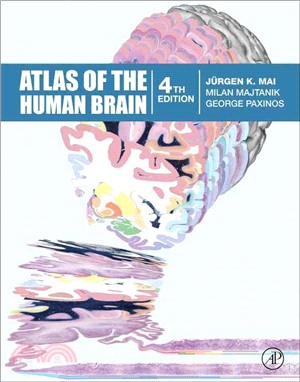
Atlas of the Human Brain
商品資訊
ISBN13:9780128028001
出版社:Academic Pr
作者:Juergen K. Mai; Milan Majtanik; George Paxinos
出版日:2015/11/30
裝訂/頁數:精裝/456頁
規格:36.8cm*28.6cm*3.2cm (高/寬/厚)
版次:4
商品簡介
The fourth edition of Atlas of the Human Brain presents the anatomy of the brain at macroscopic and microscopic levels, featuring different aspects of brain morphology and topography. This greatly enlarged new edition provides the most detailed and accurate delineations of brain structure available. It includes features which assist in the new fields of neuroscience - functional imaging, resting state imaging and tractography.Atlas of the Human Brain is an essential guide to those working with human brain imaging or attempting to relate their observations on experimental animals to humans. Totally new in this edition is the inclusion of Nissl plates with delineation of cortical areas (Brodmann’s areas), the first time that these areas have been presented in serial histological sections.
‧ The contents of the Atlas of the brain in MNI stereotaxic space has been extensively expanded from 143 pages, showing 69 levels through the hemisphere, to 314 pages representing 99 levels. ‧ In addition to the fiber-stained (myelin) plates, we now provide fifty new (Nissl) plates covering cytoarchitecture. These are interdigitated within the existing myelin plates of the stereotaxic atlas. ‧ All photographic plates now represent the complete hemisphere. ‧ All photographs of the cell- and fiber-stained sections have been transformed to fit the MNI-space. ‧ Major fiber tracts are identified in the fiber-stained sections. ‧ In the Nissl plates cortical delineations (Brodmann’s areas) are provided for the first time. ‧ The number of diagrams increased to 99. They were now generated from the 3D reconstruction of the hemisphere registered to the MNI- stereotaxic space. They can be used for immediate comparison between our atlas and experimental and clinical imaging results. ‧ Parts of cortical areas are displayed at high magnification on the facing page of full page Nissl sections. Images selected highlight those areas which are thought to correspond with those published by von Economo and Koskinas (1925). ‧ A novel way of depicting cortical areal pattern is used: The cortical cytoarchitectonic ribbon is unfolded and presented linearly. This linear representation of the cortex enables the comparison of different interpretations of cortecal areas and allows mapping of activation sites. ‧ Low magnification diagrams in the horizontal (axial) and sagittal planes are included, calculated from the 3D model of the atlas brain.
主題書展
更多書展今日66折
您曾經瀏覽過的商品
購物須知
外文書商品之書封,為出版社提供之樣本。實際出貨商品,以出版社所提供之現有版本為主。部份書籍,因出版社供應狀況特殊,匯率將依實際狀況做調整。
無庫存之商品,在您完成訂單程序之後,將以空運的方式為你下單調貨。為了縮短等待的時間,建議您將外文書與其他商品分開下單,以獲得最快的取貨速度,平均調貨時間為1~2個月。
為了保護您的權益,「三民網路書店」提供會員七日商品鑑賞期(收到商品為起始日)。
若要辦理退貨,請在商品鑑賞期內寄回,且商品必須是全新狀態與完整包裝(商品、附件、發票、隨貨贈品等)否則恕不接受退貨。


