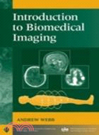
Introduction To Biomedical Imaging
商品資訊
ISBN13:9780471237662
出版社:John Wiley & Sons Inc
作者:Webb
出版日:2002/12/12
裝訂/頁數:精裝/264頁
規格:23.5cm*15.9cm*1.9cm (高/寬/厚)
定價
:NT$ 7467 元優惠價
:
90 折 6720 元
若需訂購本書,請電洽客服 02-25006600[分機130、131]。
商品簡介
作者簡介
目次
商品簡介
An integrated, comprehensive survey of biomedical imaging modalities
An important component of the recent expansion in bioengineering is the area of biomedical imaging. This book provides in-depth coverage of the field of biomedical imaging, with particular attention to an engineering viewpoint.
Suitable as both a professional reference and as a text for a one-semester course for biomedical engineers or medical technology students, Introduction to Biomedical Imaging covers the fundamentals and applications of four primary medical imaging techniques: magnetic resonance imaging, ultrasound, nuclear medicine, and X-ray/computed tomography.
Taking an accessible approach that includes any necessary mathematics and transform methods, this book provides rigorous discussions of:
The physical principles, instrumental design, data acquisition strategies, image reconstruction techniques, and clinical applications of each modality
Recent developments such as multi-slice spiral computed tomography, harmonic and sub-harmonic ultrasonic imaging, multi-slice PET scanning, and functional magnetic resonance imaging
General image characteristics such as spatial resolution and signal-to-noise, common to all of the imaging modalities
作者簡介
ANDREW WEBB, PhD, is a faculty member in the Department of Electrical and Computer Engineering and the Beckman Institute for Advanced Science and Technology at the University of Illinois at Urbana-Champaign. Dr. Webb has contributed to many areas of magnetic resonance imaging including developments in radiofrequency coil design, feedback control of thermal processes, techniques for localized spectroscopy, and functional brain mapping. He was awarded a Whitaker Foundation Research Award and a National Science Foundation Career Award in 1997, a Wolfgang-Paul Prize from the Alexander von Humbolt Foundation in 2001, and Xerox and Willett awards for young faculty in 2002. He is a Senior Member of the IEEE.
目次
Preface.
Acknowledgments.
1. X-Ray Imaging and Computed Tomography.
1.1 General Principles of Imaging with X-Rays.
1.2 X-Ray Production.
1.3 Interactions of X-Rays with Tissue.
1.4 Linear and Mass Attenuation Coefficients of X-Rays in Tissue.
1.5 Instrumentation for Planar X-Ray Imaging.
1.6 X-Ray Image Characteristics.
1.7 X-Ray Contrast Agents.
1.8 X-Ray Imaging Methods.
1.9 Clinical Applications of X-Ray Imaging.
1.10 Computed Tomography.
1.11 Image Processing for Computed Tomography.
1.12 Spiral/Helical Computed Tomography.
1.13 Multislice Spiral Computed Tomography.
1.14 Radiation Dose.
1.15 Clinical Applications of Computed Tomography.
2. Nuclear Medicine.
2.1 General Principles of Nuclear Medicine.
2.2 Radioactivity.
2.3 The Production of Radionuclides.
2.4 Types of Radioactive Decay.
2.5 The Technetium Generator.
2.6 The Biodistribution of Technetium-Based Agents within the Body.
2.7 Instrumentation: The Gamma Camera.
2.8 Image Characteristics.
2.9 Single Photon Emission Computed Tomography.
2.10 Clinical Applications of Nuclear Medicine.
2.11 Positron Emission Tomography.
3. Ultrasonic Imaging.
3.1 General Principles of Ultrasonic Imaging.
3.2 Wave Propagation and Characteristic Acoustic Impedance.
3.3 Wave Reflection and Refraction.
3.4 Energy Loss Mechanisms in Tissue.
3.5 Instrumentation.
3.6 Diagnostic Scanning Modes.
3.7 Artifacts in Ultrasonic Imaging.
3.8 Image Characteristics.
3.9 Compound Imaging.
3.10 Blood Velocity Measurements Using Ultrasound.
3.11 Ultrasound Contrast Agents, Harmonic Imaging, and Pulse Inversion Techniques.
3.12 Safety and Bioeffects in Ultrasonic Imaging.
3.13 Clinical Applications of Ultrasound.
4. Magnetic Resonance Imaging.
4.1 General Principles of Magnetic Resonance Imaging.
4.2 Nuclear Magnetism.
4.3 Magnetic Resonance Imaging.
4.4 Instrumentation.
4.5 Imaging Sequences.
4.6 Image Characteristics.
4.7 MRI Contrast Agents.
4.8 Magnetic Resonance Angiography.
4.9 Diffusion-Weighted Imaging.
4.10 In Vivo Localized Spectroscopy.
4.11 Functional MRI.
4.12 Clinical Applications of MRI.
5. General Image Characteristics.
5.1 Introduction.
5.2 Spatial Resolution.
5.3 Signal-to-Noise Ratio.
5.4 Contrast-to-Noise Ratio.
5.5 Image Filtering.
5.6 The Receiver Operating Curve.
Appendix A: The Fourier Transform.
Appendix B: Backprojection and Filtered Backprojection.
Abbreviations.
Index.
主題書展
更多
主題書展
更多書展購物須知
外文書商品之書封,為出版社提供之樣本。實際出貨商品,以出版社所提供之現有版本為主。部份書籍,因出版社供應狀況特殊,匯率將依實際狀況做調整。
無庫存之商品,在您完成訂單程序之後,將以空運的方式為你下單調貨。為了縮短等待的時間,建議您將外文書與其他商品分開下單,以獲得最快的取貨速度,平均調貨時間為1~2個月。
為了保護您的權益,「三民網路書店」提供會員七日商品鑑賞期(收到商品為起始日)。
若要辦理退貨,請在商品鑑賞期內寄回,且商品必須是全新狀態與完整包裝(商品、附件、發票、隨貨贈品等)否則恕不接受退貨。


